| Table of Contents | |
|
Case Report
| ||||||
| Scar sarcoidosis: A case report | ||||||
| Gayathri Devi HJ1, Shivaswamy KN2, Rashmi Krishnappa3 | ||||||
|
1Department of Respiratory Medicine, MS Ramaiah Medical College, Bangalore, India.
2Department of Dermatology, MS Ramaiah Medical College, Bangalore, India. 3Department of Pathology, MS Ramaiah Medical College, Bangalore, India. | ||||||
| ||||||
|
[HTML Abstract]
[PDF Full Text]
[Print This Article]
[Similar article in Pumed] [Similar article in Google Scholar] |
| How to cite this article |
| Gayathri Devi HJ, Shivaswamy KN, Krishnappa R. Scar sarcoidosis: A case report. Edorium J Respir Med 2014;1:1–4. |
|
Abstract
|
|
Sarcoidosis is an enigmatic disease with multisystem involvement. Cutaneous manifestations of sarcoidosis are relatively uncommon and scar sarcoidosis is rare. We report a case of scar sarcoidosis in a 53-year-old female who developed a nodular lesion at the nasal bridge before developing pulmonary manifestations. A short course of oral steroids caused regression of the lesion.
| |
|
Keywords:
Cutaneous, Granuloma, Sarcoidosis, Scar
| |
|
Introduction
| ||||||
|
Sarcoidosis is a multi-system disease of unknown aetiology characterized by the formation of non-caseating granulomas in the affected organs. Involvement of the skin in sarcoidosis occurs in 25 % of cases [1]. Infiltration of sarcoid granuloma in old scars is one of the uncommon cutaneous manifestations of sarcoidosis [2]. We report a case of scar sarcoidosis in a 53-year-old female who developed a nodular lesion over the 20 years old scar before developing pulmonary manifestations. | ||||||
|
Case Report
| ||||||
|
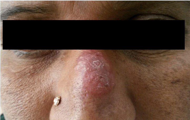
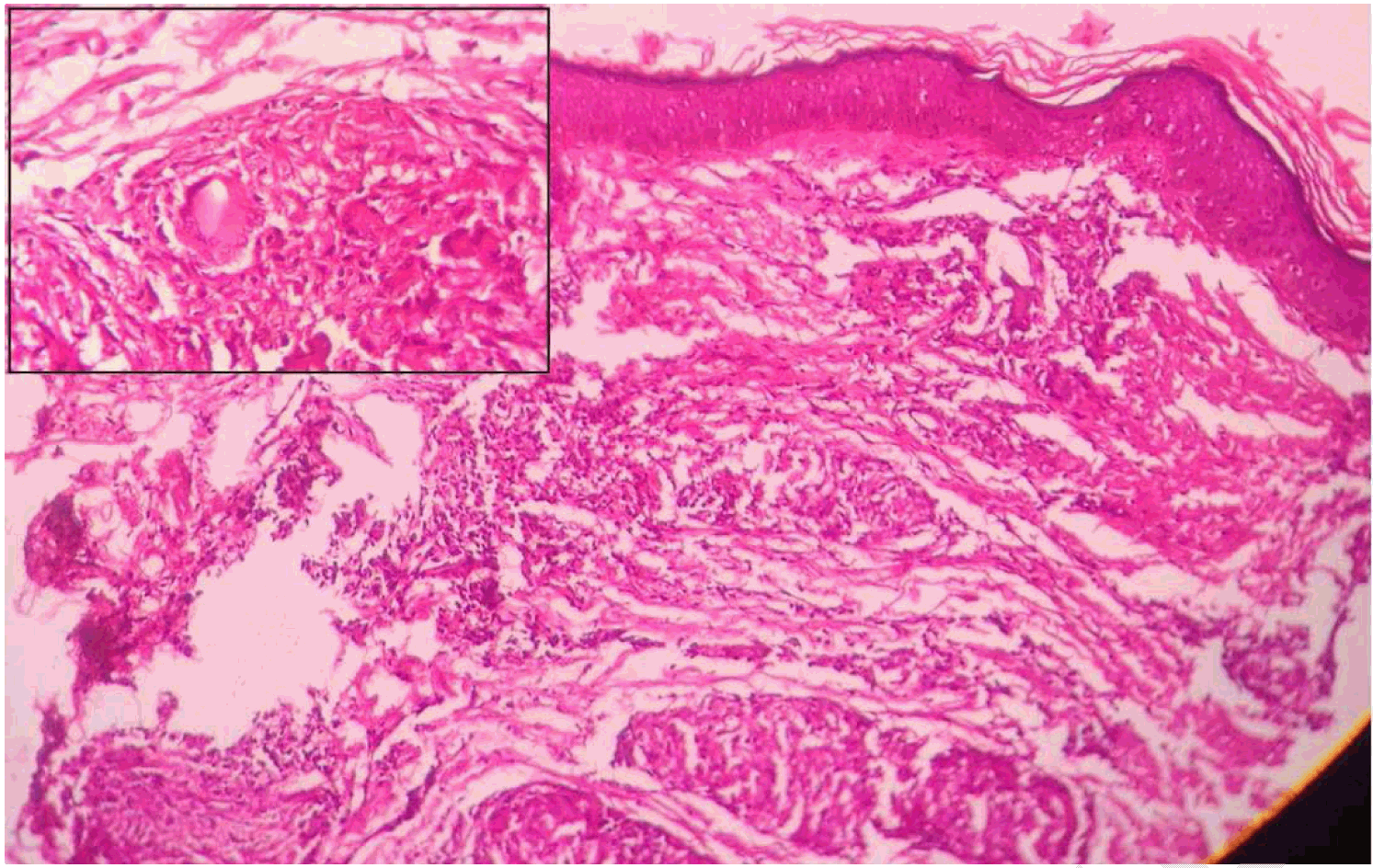
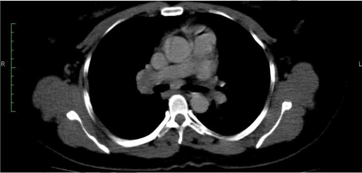
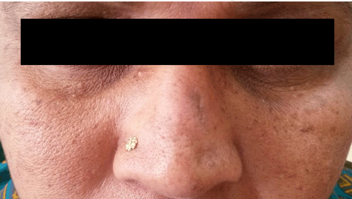
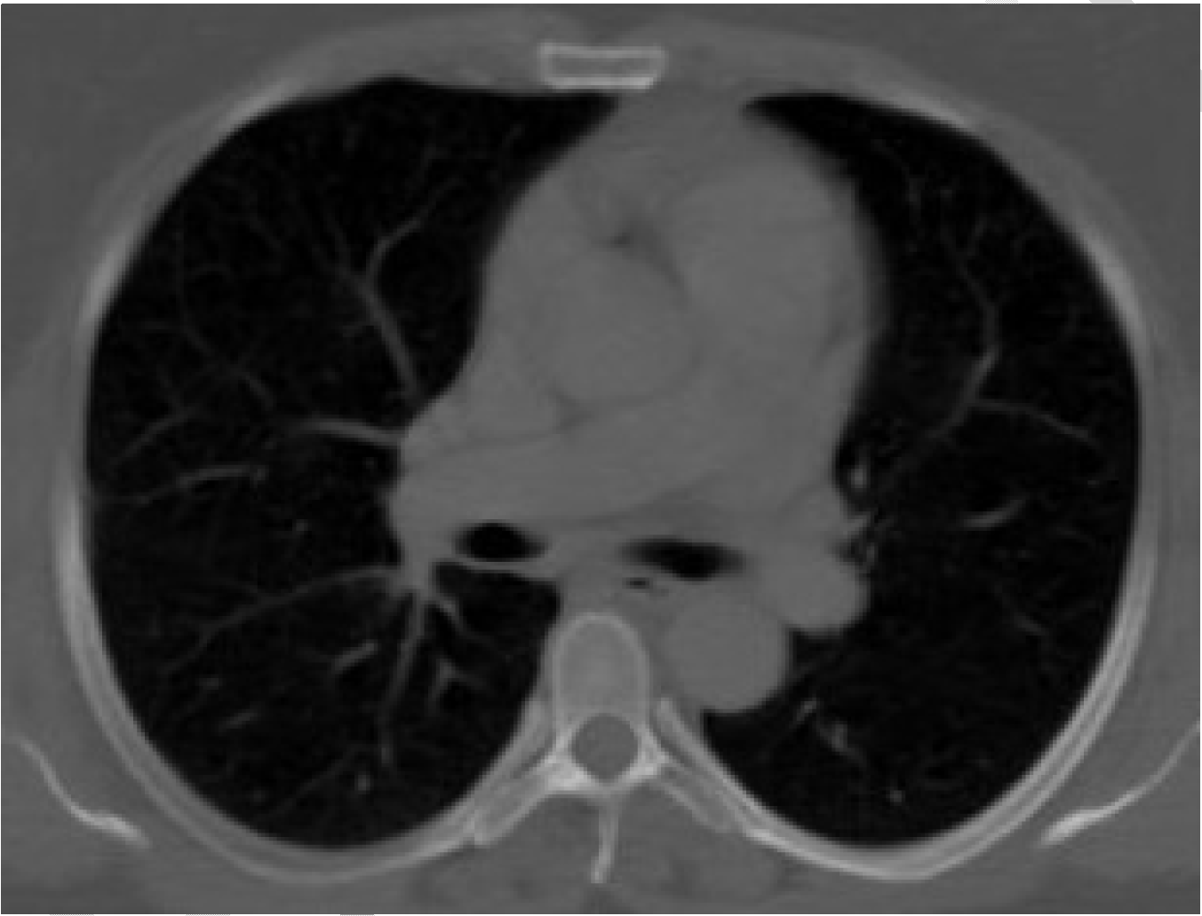
A 53-year-old female presented to the dermatology department with complaints of swelling over the nasal bridge for two months. It started as a small nodule and increased in size over two months (Figure 1). She gave a history of trauma to nose 20 years back which had left a scar. On examination, she had a firm swelling over the nasal bridge measuring 2.5x2 cm. There was no lymphadenopathy. Systemic examination was normal. Mantoux test was negative. Routine hematological investigations were normal. The serum ACE level was elevated 88 IU/L (18–55 IU/L). Biopsy of the nodule showed chronic granulomatous lesion suggestive of sarcoidosis (Figure 2)). The patient received three doses of intralesional steroid injection without any relief. She was referred to respiratory medicine department for cough of five days duration, six months after her first presentation to dermatology department. She gave a history of exertional breathlessness of six months duration. Clinical examination was normal. Chest X-ray showed right hilar prominence for which patient was subjected to contrast enhanced computed tomography (CECT) thorax, which revealed mediastinal lymphadenopathy without any parenchymal involvement (Figure 3). Further workup did not reveal any ophthalmologic, neurologic, cardiovascular or renal involvement. Ultrasound of the abdomen, 2D echocardiogram and spirometry were normal. The patient was started on oral prednisolone 30 mg per day for three months, then tapered over the next three months and stopped. Treatment resulted in complete regression of the skin lesion (Figure 4). CECT of thorax after six months of treatment was normal (Figure 5). | ||||||
| ||||||
| ||||||
| ||||||
| ||||||
| ||||||
|
Discussion
| ||||||
|
Sarcoidosis is a systemic granulomatous disease that affects most commonly the lungs, hilar and mediastinal lymph nodes or both. It may involve any organ in the body and may present with various clinical manifestations. Dermatologic manifestations are relatively uncommon and are seen in 25% of patients with sarcoidosis. It occurs at all ages, although usually develops before 50 years with peak incidence at 20–39 years [3]. Skin sarcoid was first described by Caesar Boek in 1899 [4]. Cutaneous manifestations of sarcoidosis could be specific lesions which show non-caseating granuloma on biopsy or non-specific lesions which do not have granulomas [1]. Cutaneous sarcoidosis is known as one of the 'great imitators' in dermatology because of different morphologies of the lesions [5]. Skin manifestations include erythema nodosum, lupus pernio, papules, nodules and plaques. Scar sarcoidosis is a form of cutaneous sarcoidosis [6]. It refers to lesions of cutaneous sarcoidosis that appear in pre-existing scars [7]. Old scars are infiltrated with non-caseating epithelioid cell granulomas. Islands of epithelioid cells may have a few Langerhans giant cells. Giant cells may contain asteroid or Schaumann bodies. Granulomas are usually in the superficial dermis but may involve the full thickness of the dermis and extend into the subcutaneous tissue [1]. Granulomas are referred to as naked because they have only a sparse lymphocytic infiltrate at the margins of the granulomas [7]. Scar sarcoidosis can occur in scars from previous wounds, tattoo scars, site of intramuscular injection and herpes zoster scar. The pathogenesis has been hypothesized to be due to hypersensitivity reaction or foreign body contamination of the scar [8]. It can occur in varying intervals. Old scar reactivation may be the only manifestation of cutaneous sarcoidosis [9]. It may precede or accompany systemic involvement [10]. Skin lesions may be the only manifestation of sarcoidosis and may guide an appropriate diagnosis since they are an easily accessible source of tissue for histologic diagnosis [7]. Sarcoidal granulomas produce angiotensin converting enzyme and elevated levels are found in 60% of patients with sarcoidosis [3]. Sensitivity of the test is 60% and specificity is 70%.There may be decrease in the levels of serum ACE in response to therapy [11]. However, decision to treat is not based on the serum ACE level. Studies have shown that the other markers like soluble IL-2 receptor (SIL-2R) level and chitotriosidase are increased in pulmonary and extrapulmonary sarcoidosis. Krebs Von Den Lungen-6 (KL-6) is a mucin produced by pneumocytes type II and cells of the bronchial epithelium. Increased levels of this marker have been reported in the fibrosis form of pulmonary sarcoidosis [12][13]. Scar sarcoidosis case needs a complete evaluation for systemic involvement as in our case. The treatment of cutaneous sarcoidosis still remains a challenge [14]. Spontaneous remission of the lesion is seen in up to two-thirds of patients [15] . Treatment of cutaneous sarcoidosis includes both local and systemic therapies. During mild limited skin disease treatment is with topical corticosteroid or repeated intralesional injections of triamcinolone. In patients requiring systemic therapy for recalcitrant or deforming skin lesions, corticosteroids alone or with antimalarials or methotrexate may be indicated [16]. Drug of first choice remains oral corticosteroids with a daily dose of 30–40 mg of prednisolone for 6–12 weeks. Based on the clinical response doses are reduced and continued for 6–12 months. Methotrexate in the dose of 7.5–25 mg weekly has also been used to treat systemic and cutaneous sarcoidosis. The above mentioned treatments often result in an incomplete clinical response or adverse events. In such situations tumor necrosis factor -alpha inhibitors, infliximab and adalimumab have been tried [16] [17]. Limited data report the use of thalidomide for patients with cutaneous sarcoidosis who are resistant to corticosteroids [18]. Laser therapy is a newer modality of treatment that may provide a quick and non-invasive treatment option for cutaneous sarcoidosis [16] . | ||||||
|
Conclusion
| ||||||
|
Patients with any form of lesion over pre-existing scars should be properly evaluated for sarcoidosis to prevent morbidity due to systemic involvement. | ||||||
|
Key Messages
| ||||||
|
We report a case of cutaneous scar sarcoidosis in a 53-year-old female patient who developed a nodular lesion in a post-traumatic scar sustained 20 years ago over the bridge of the nose. Biopsy showed sarcoid granuloma. The nodular lesion regressed with oral prednisolone treatment. | ||||||
|
References
| ||||||
| ||||||
|
[HTML Abstract]
[PDF Full Text]
|
|
Author Contributions
Gayathri Devi HJ – Substantial contributions to conception and design, Acquisition of data, Drafting the article, revising it critically for important intellectual content, Final approval of the version to be published Shivaswamy KN – Substantial contributions to conception and design, Analysis and interpretation of data, Drafting the article, Final approval of the version to be Published Rashmi Krishnappa – Group 1 - Substantial contributions to conception and design, Drafting the article, revising it critically for important intellectual content, Final approval of the version to be published |
|
Guarantor of submission
The corresponding author is the guarantor of submission. |
|
Source of support
None |
|
Conflict of interest
Authors declare no conflict of interest. |
|
Copyright
© 2014 Gayathri Devi HJ et al. This article is distributed under the terms of Creative Commons Attribution License which permits unrestricted use, distribution and reproduction in any medium provided the original author(s) and original publisher are properly credited. Please see the copyright policy on the journal website for more information. |
|
|